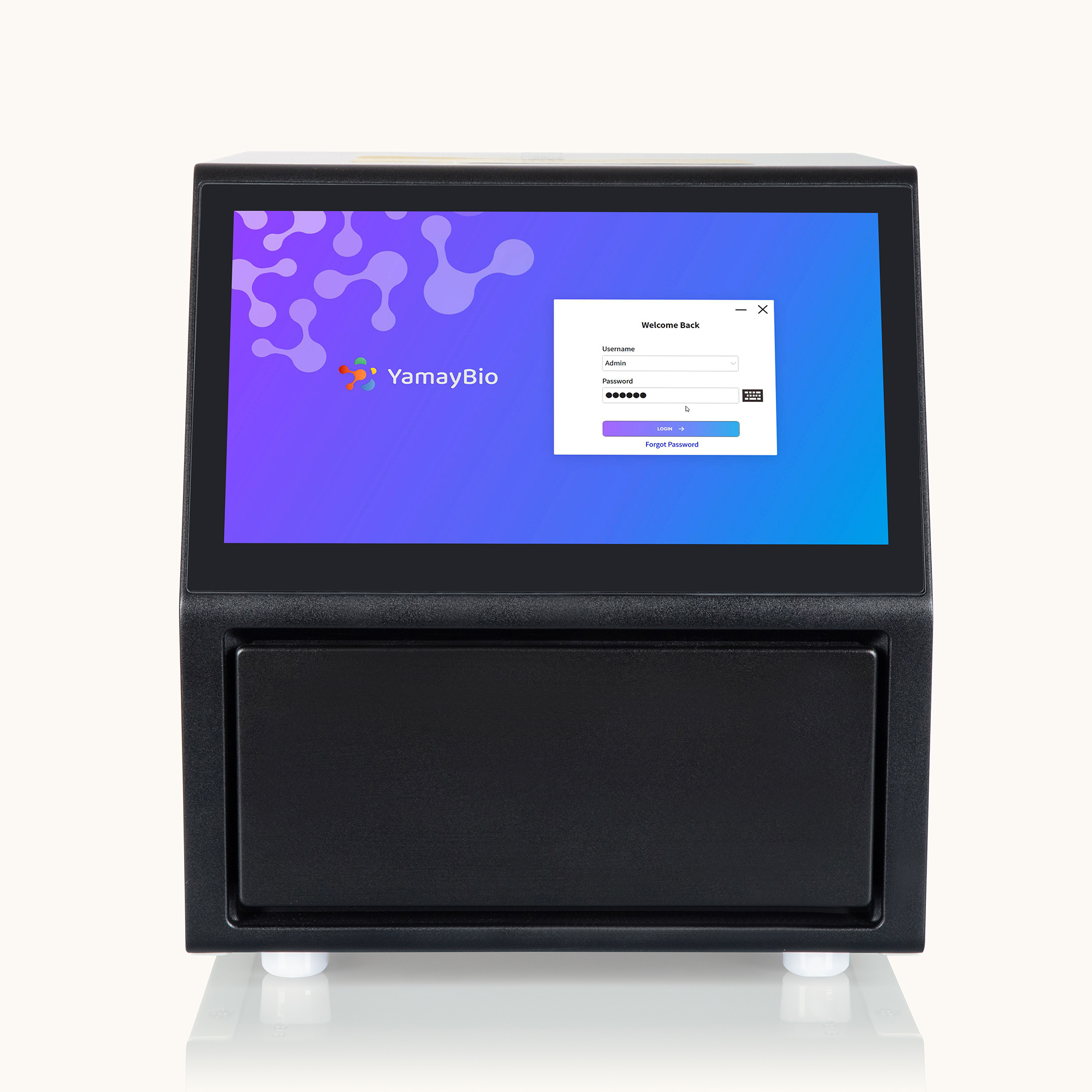
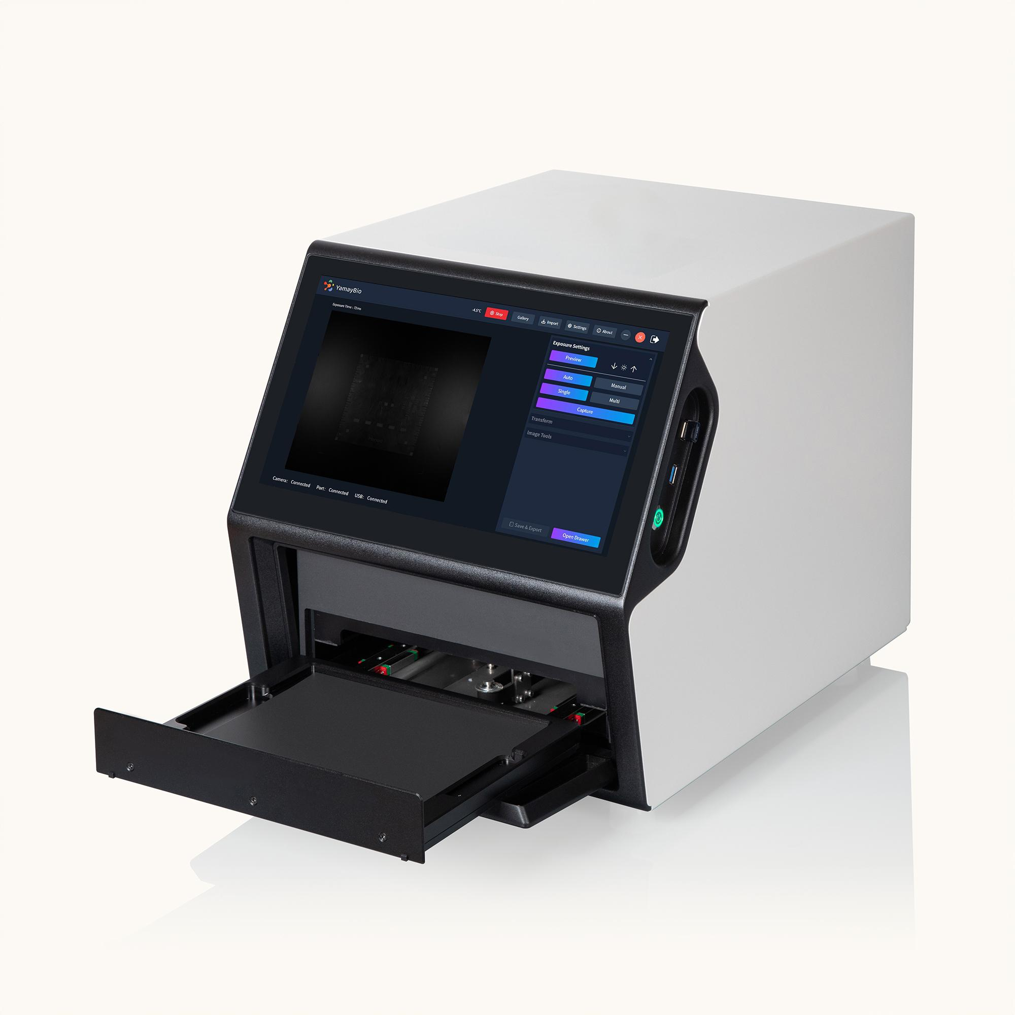

SignalView CL Imaging System
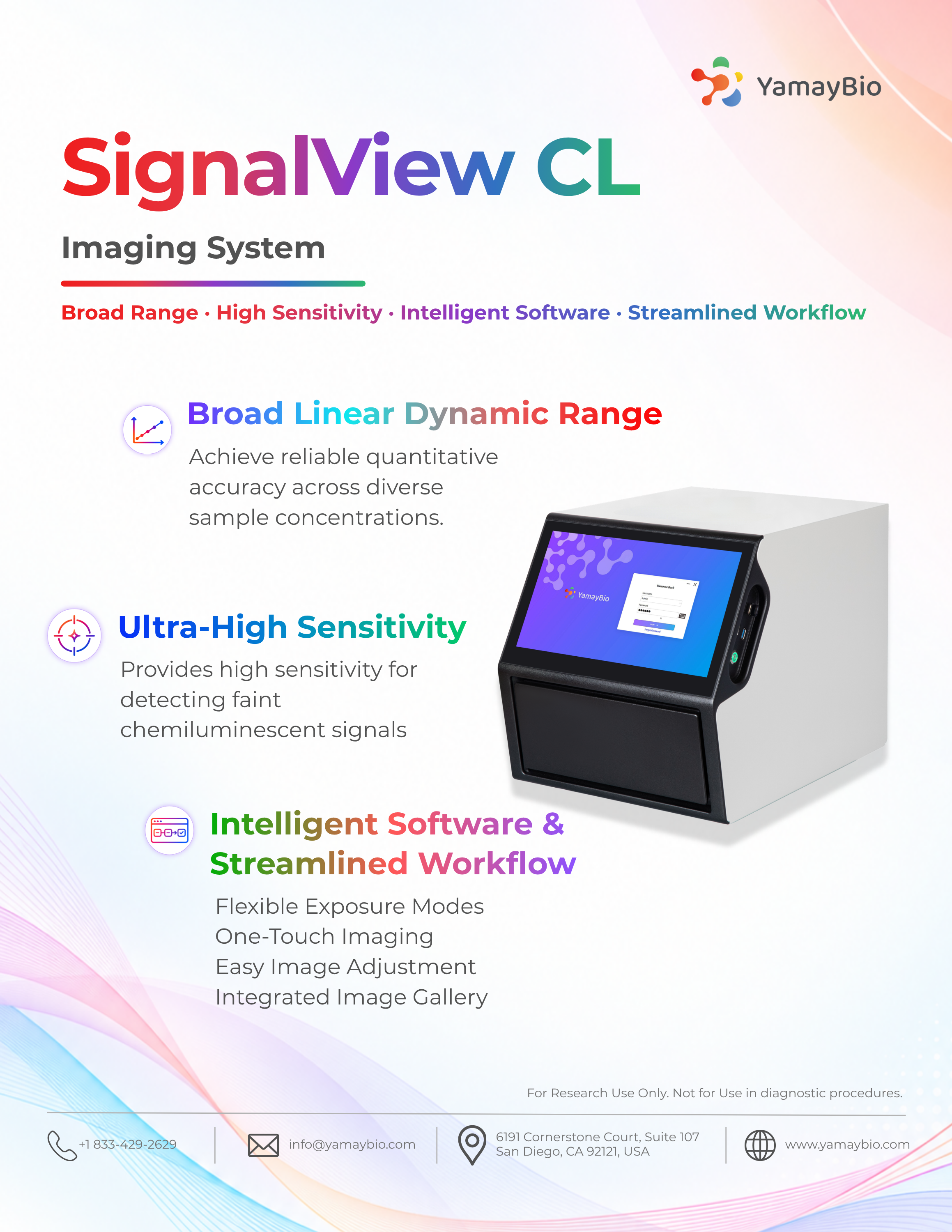
The SignalView CL Imaging System is engineered for sensitive and reliable chemiluminescence imaging. Its Scientific CMOS (sCMOS) detector combines high quantum efficiency with low read noise to improve weak-signal detection, while four-stage Peltier cooling reduces dark current and thermal noise for cleaner backgrounds and stable performance during long exposures. With a broad linear detection range, high sensitivity, and intuitive software, SignalView CL supports accurate quantification and streamlines chemiluminescence imaging to improve workflow efficiency and consistency from image capture to analysis. Read more
Interested in this product? Contact us for a demo, pricing, and more information.
Instructional Video: https://www.youtube.com/watch?v=3RVuyR41gCo&t=2s
The SignalView CL Imaging System is engineered for sensitive and reliable chemiluminescence imaging. Its Scientific CMOS (sCMOS) detector combines high quantum efficiency with low read noise to improve weak-signal detection, while four-stage Peltier cooling reduces dark current and thermal noise for cleaner backgrounds and stable performance during long exposures. With a broad linear detection range, high sensitivity, and intuitive software, SignalView CL supports accurate quantification and streamlines chemiluminescence imaging to improve workflow efficiency and consistency from image capture to analysis.
KEY FEATURES

Broad Linear Dynamic Range

Ultra-High Sensitivity

Intelligent Software & Streamlined Workflow
Broad Linear Dynamic Range
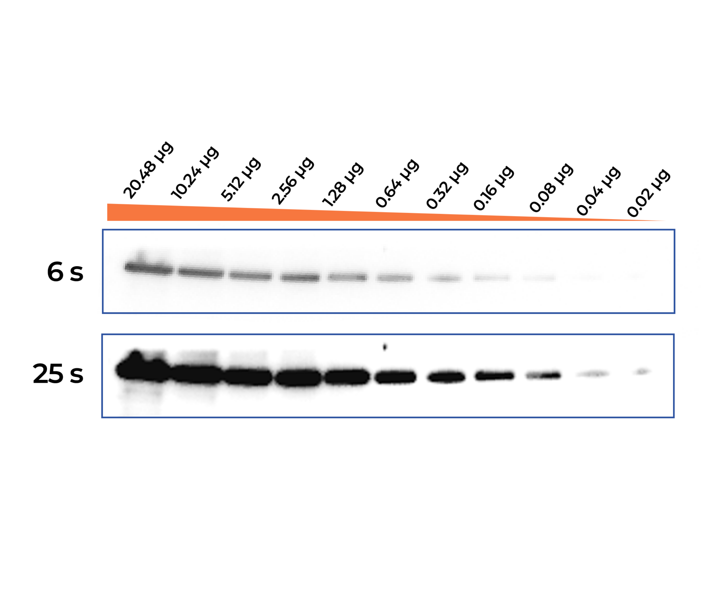
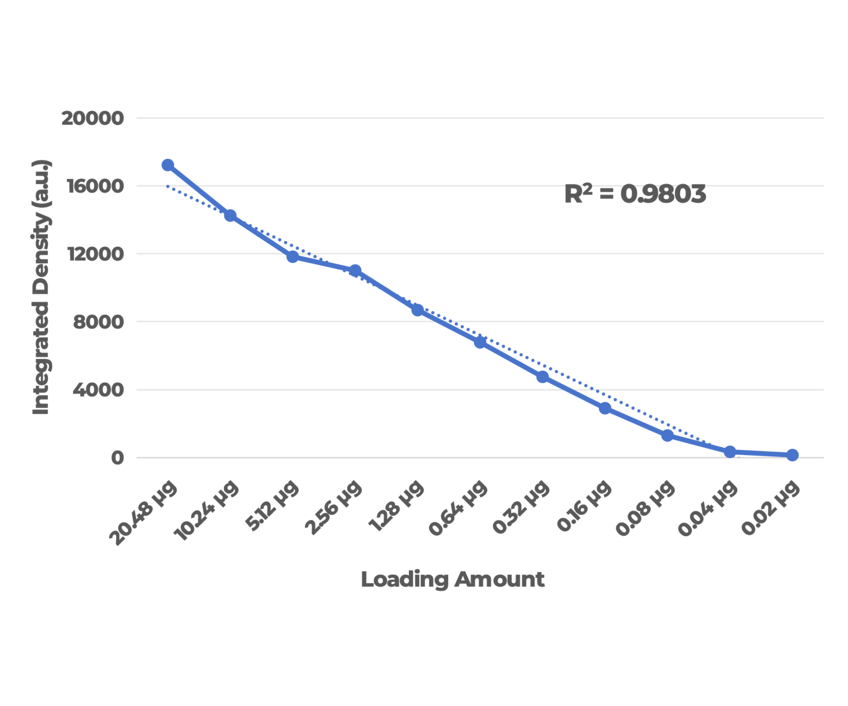
Achieve advanced quantitative accuracy across diverse sample concentrations. The system exhibits an outstanding linear correlation across a nearly 1000-fold serial dilution range (20.48 to 0.02 μg). This broad linear range helps minimize signal saturation and supports reliable Western blot densitometry and relative quantification.
Ultra-High Sensitivity
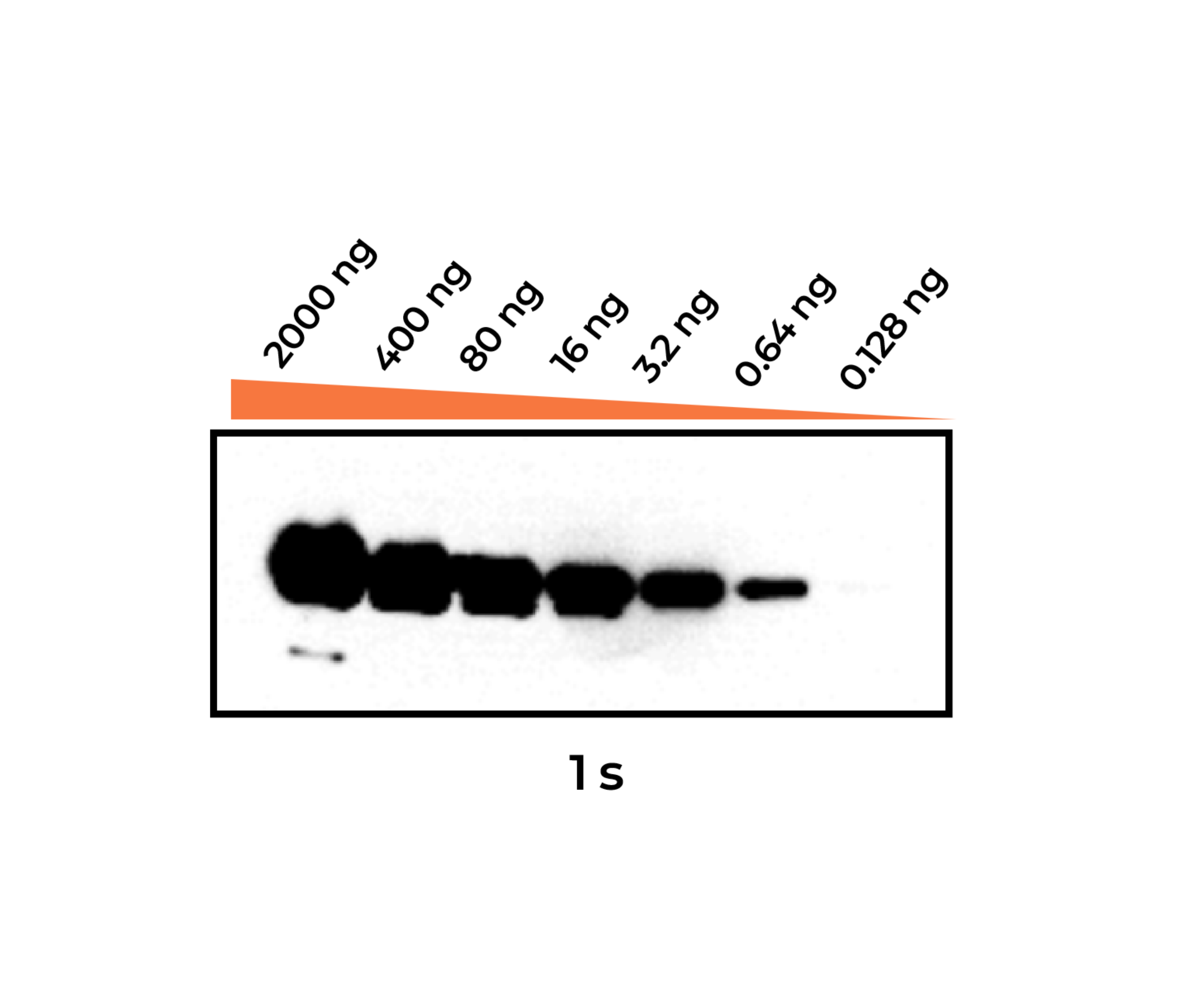
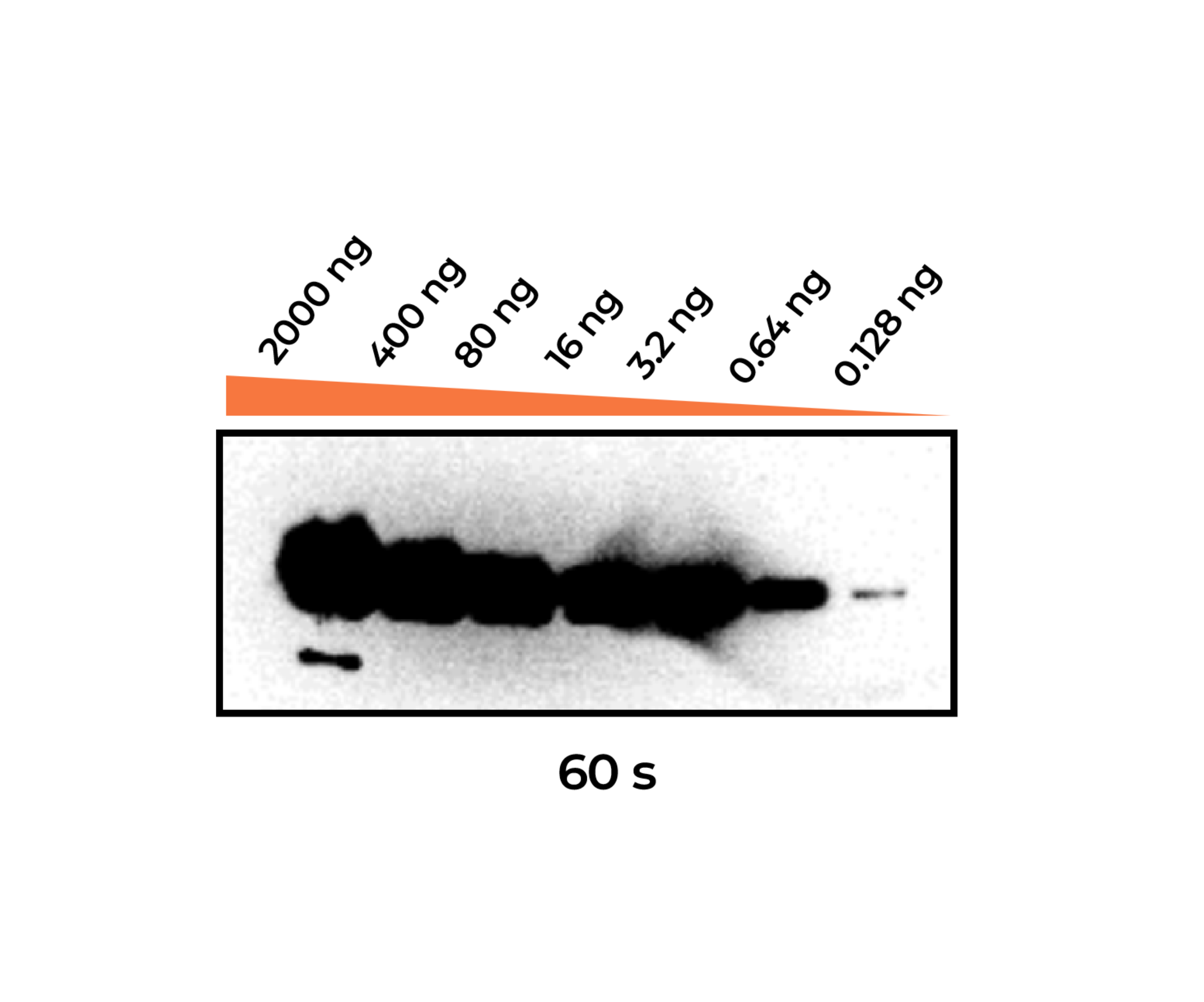
SignalView CL provides high sensitivity for detecting faint chemiluminescent signals. Distinct target bands remain visible at loading amounts as low as 0.02 μg for cell lysates (see Figure 1) and 0.128 ng for recombinant proteins (see Figure 2), supporting reliable detection of low-abundance targets.
Recombinant GFP-FLAG Protein Serial Dilution Assay. Proteins were separated on a 12% Bis-Tris precast gel with serial dilutions from 2000 to 0.128 ng per lane. Proteins were detected using an HRP-conjugated mouse anti-FLAG antibody. Blots were imaged following exposure at the indicated exposure times, with distinct bands observable even at the lowest loading amount of 0.128 ng with a 60-second exposure.
Intelligent Software & Streamlined Workflow
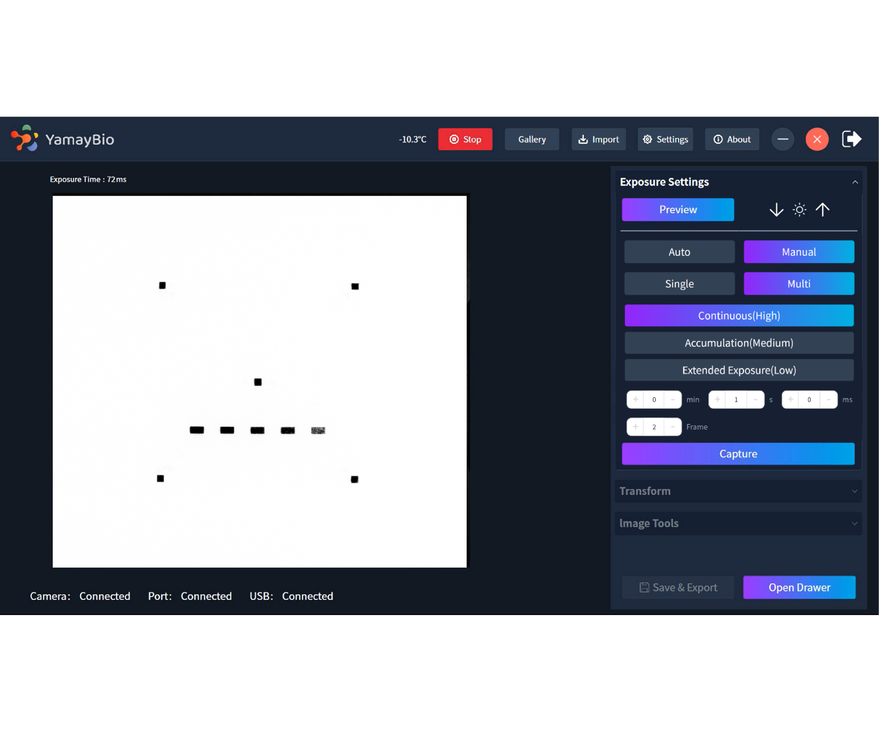
Flexible Exposure Modes: Single and multiple exposure options with automatic or manual control for imaging both strong and weak signals.
One-Touch Imaging: Capture chemiluminescence, marker, and overlay images in a streamlined workflow.
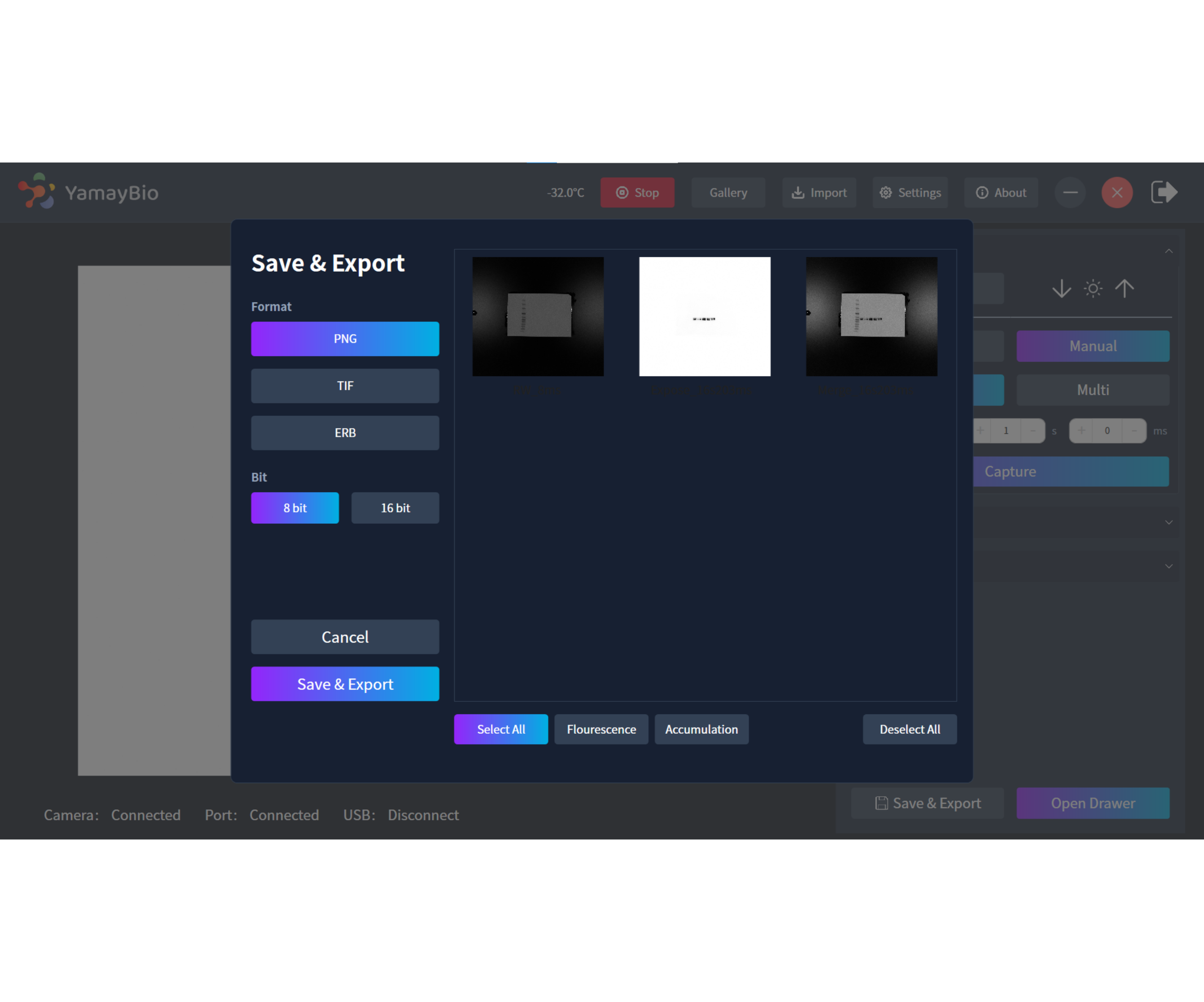
Batch Export: Export multiple images in one step.
Easy Image Adjustment: Quickly rotate and adjust images with intuitive controls.
Original Image Access: Save and reopen original image files at any time.
Related Products

Product Manual



Request a Demo

Specifications
| Display | 13.3-inch HD Touchscreen (1920×1080) |
| Detector / Camera | 15 MP sCMOS (4/3 inch) |
| Pixel Performance | 3.8 × 3.8 μm pixel, 95% QE, 1.0 e- RMS noise |
| Cooling | Four-stage Peltier (-30°C @ 25°C ambient) |
| Optics | 25 mm F0.95 lens |
| Imaging | 17 × 19 cm area (2×2, 4×4, 8×8 binning) |
| Dimensions (W x D x H) | 33.1 × 45.0 × 37 cm (13.0 × 17.7 × 14.6 in) |
| Weight | 24.2 kg (53.4 lb) |
| Power Supply | AC 80–264 V, 47–63 Hz, 200 VA MAX |