


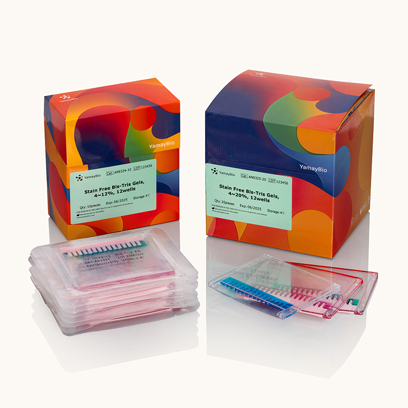
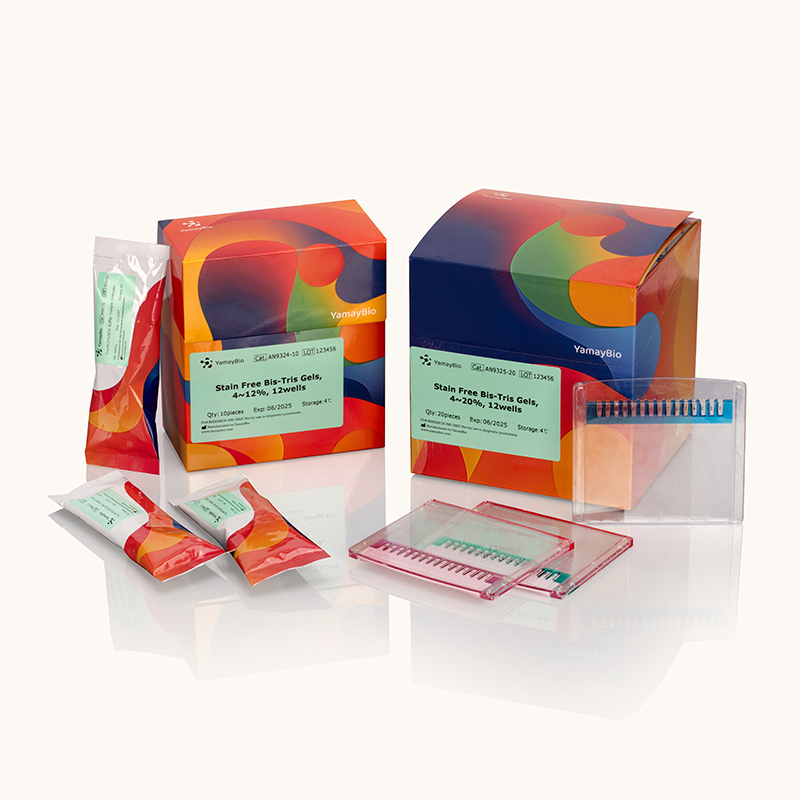
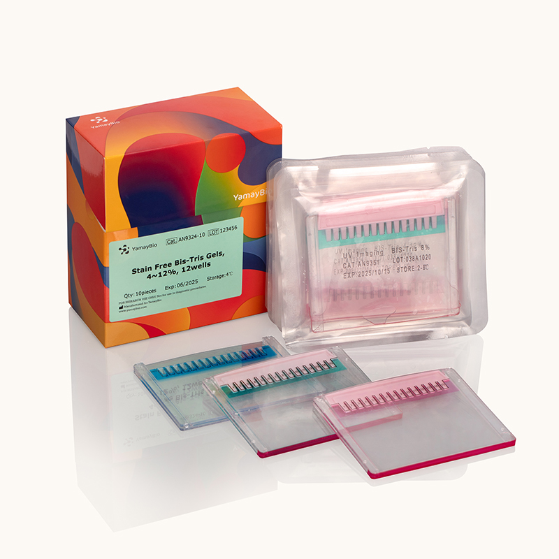
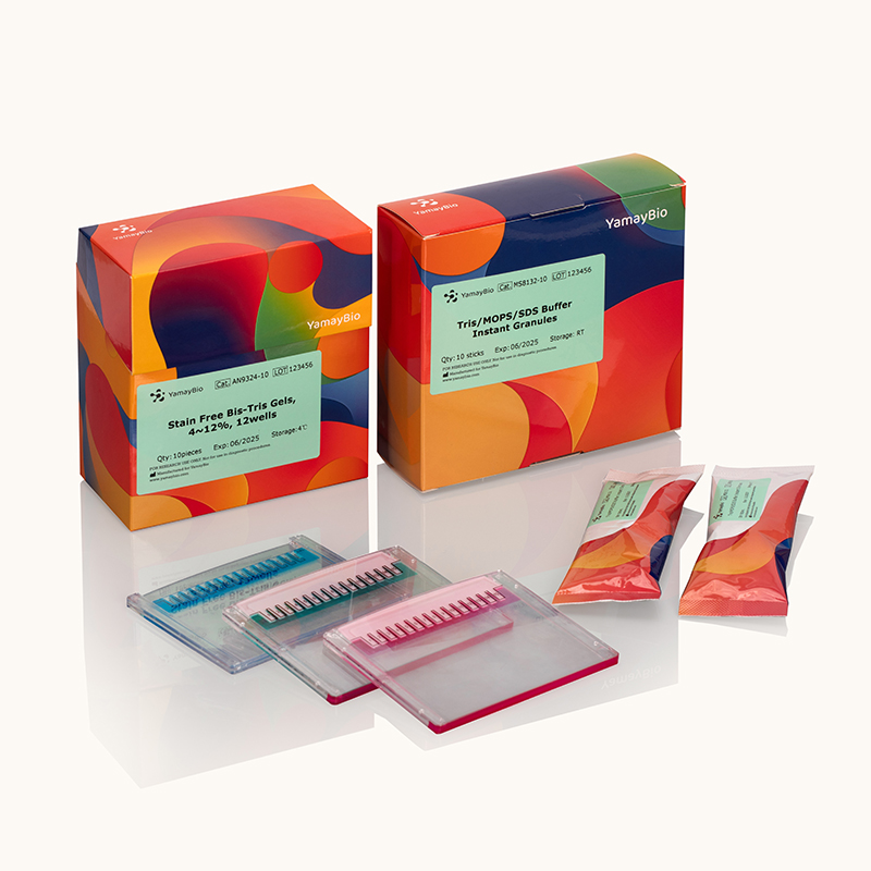
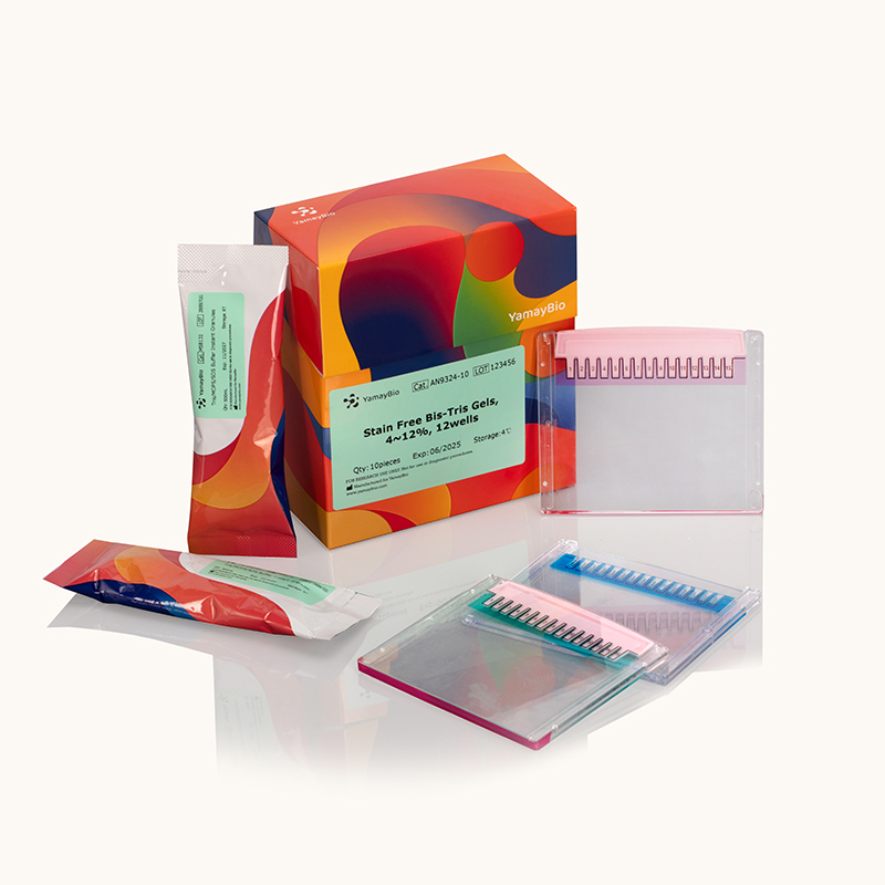


Stain-Free Bis-Tris Precast Gels
Stain-Free Bis-Tris Precast Gels are a safe, fast, and high-performance precast polyacrylamide gel designed for protein separation. Read more
Transport at room temperature and Store at 4°C. Do not freeze.
Stain-Free Bis-Tris precast gels are a safe, fast, and high-performance precast polyacrylamide gel system designed for protein separation. Protein bands on these gels can be directly imaged under UV light, eliminating the need for staining. Our precast gels are available in 12 well or 15 well formats, with recommended maximum sample volumes of 35 μL and 25 μL, respectively. The dimensions are as follows:
Gel plate: 100 × 85 × 4.7 mm
Gel: 85 × 70 × 1 mm
Additionally, the Tris/MOPS/SDS electrophoresis buffer provided with the gels is neutral, which enhances gel stability and prevents protein re-modification during electrophoresis.
This product can be transported at room temperature but should not be exposed to temperatures below 0°C. It should be stored at 4°C, with a shelf life of 12 months under proper storage conditions. Do not freeze the Stain Free Bis-Tris precast gels, as use of expired or improperly stored gels may result in poor band resolution.
KEY FEATURES

Colorful Stacking Gels Easy sample loading.

Neutral pH Resulting in sharper band resolution and accurate results.

Stain Free Protein bands can be observed after electrophoresis and transfer without additional staining.
Non-migratory
The dyes do not migrate during electrophoresis.
The dyes do not diffuse or fade during gel staining and destaining.
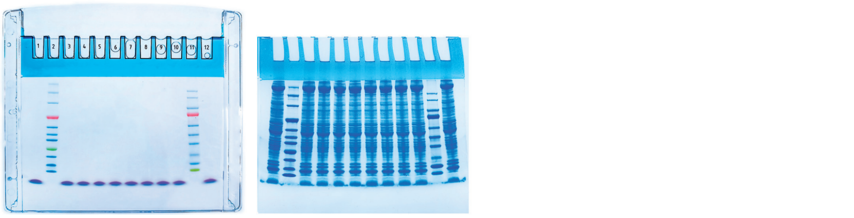
Stain-Free
Gels enable the direct visualization of protein bands under UV light both before and after transfer to a membrane.
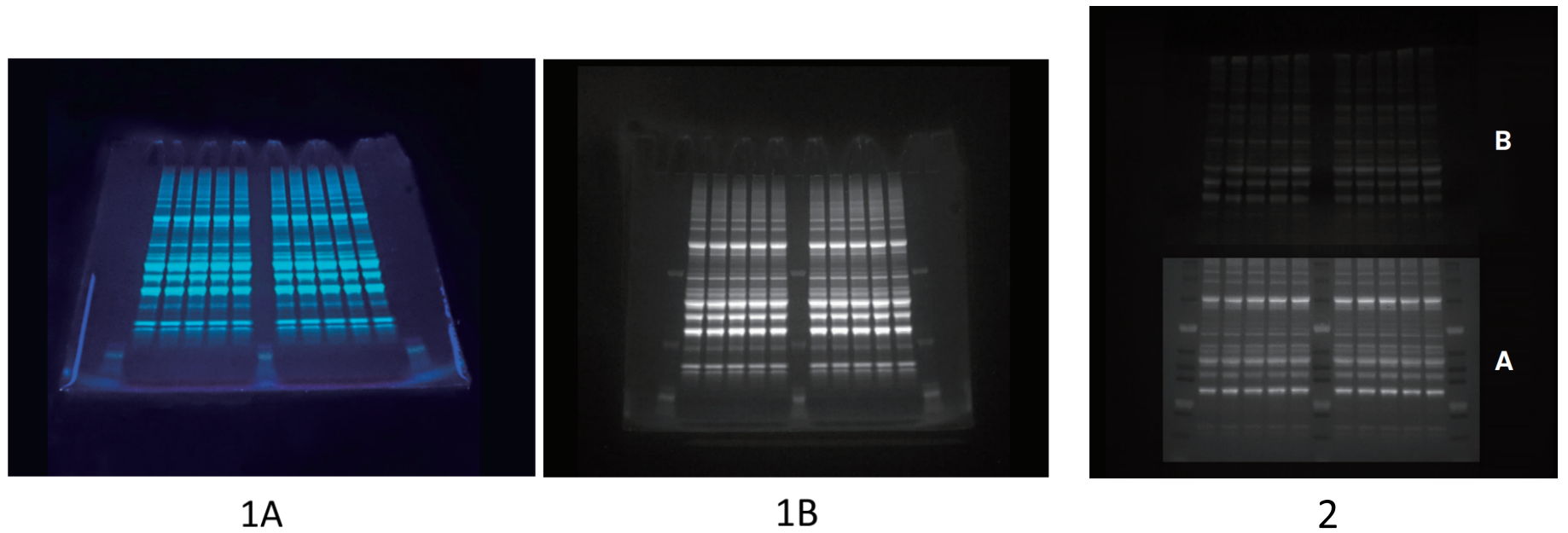
1A: Stain-free visualization of proteins in gel, imaging at UV 302nm, taken by mobile phone;
1B: Stain-free visualization of proteins in gel with stain-free enabled imaging systems;
2: UV images of A. post-transfer PVDF membrane; B. protein gel.
Related Products

Protein Electrophoresis
Stain-Free Gradient-like PAGE Gel Kit
Product Code: RF1541

Protein Electrophoresis
One-Step Stain-Free PAGE Gel Rapid Preparation Kit
Product Code: RF1521
Protein Electrophoresis
All-in-One Low Range Protein Gel Kit
Product Code: RF1531
Protein Electrophoresis
Tricolor Prestained Protein Ladder 10-180 kDa (Orange, Green & Blue)
Product Code: CY5463
Protein Electrophoresis
Tricolor Prestained Protein Ladder 10-250 kDa (Orange, Green & Blue)
Product Code: CY5466
Protein Electrophoresis
Protein Sample Loading Buffer (Denaturing, non-Reducing, 5×)
Product Code: GB4354
Protein Electrophoresis
Fast-dissolving Dual-color Protein Loading Buffer (Denaturing, Reducing, 5×)
Product Code: GB4353
Certificate of Analysis
Batch number
Certificate type
Date
Catalog Number(s)
Controls


 Show more
Safety Data Sheet
Show more
Product Manual

FAQs
Q1 : I have set up my gel to run and have switched on the power supply, but my gel is not running and there is no voltage or current reading on the power supply. What is wrong?
A : 1. Double check that the tape on the bottom of the gel has been removed.
2. Make sure that the gel(s) are oriented so that the taller sides of the cassette (with the printing) are facing the outside of the electrophoresis unit.
3. Make sure that the inner buffer chamber is filled sufficiently so that the wells are covered with buffer. If the wells are not covered, check for leaks and reseal.
4. Double check to see if there are any loose electrodes or connections on the Mini cell unit.
5. Check the power supply unit.

Q2 : I accidentally froze my gels. Is it still okay to use them?
A : Not recommend. They may have become brittle and may crack either before or after the run, or even when in the staining solution. May cause the formation of bubbles during the run and band distortion particularly in the center lanes.
Q3 : Working principle of stain-free gels
A : It involves incorporating a trihalo compound into the gel matrix. Upon UV activation, it covalently binds to tryptophan residues in proteins, generating a strong fluorescent product in situ, allowing direct imaging without the need for staining.
Q4 : How to use stain-free gel?
A : After electrophoresis, remove the gel from the glass/plastic plates, moisten it with water (to prevent the gel from shrinking or curling due to dehydration during UV excitation), then place it directly on the black imaging platform for UV activation (1–5 min), and adjust to an appropriate exposure time for imaging.
Q5 : Stain-free gels can still be subjected to silver staining or Coomassie brilliant blue staining after UV excitation.
A : Yes
Q6 : The results obtained with stain-free gels are not identical to those from Coomassie brilliant blue staining.
A : Because the two methods rely on different detection principles. Stain-free gels detect tryptophan residues in proteins upon UV activation.
Coomassie brilliant blue binds to proteins through electrostatic and hydrophobic interactions, reflecting total protein amount.
Therefore, proteins with low tryptophan content may appear weak or undetectable in stain-free gels, but can be clearly visualized by Coomassie staining.
As a result, band intensity and detectability differ between the two methods.
Q7 : Bands in the outer lanes of the gel are distorted/skewed.
A : A common issue caused by edge effects, uneven temperature distribution, or inconsistent ionic strength. The outermost 1–2 lanes of the gel were left unloaded. Reducing the voltage can alleviate this problem.
Specifications
| For Use With (Equipment) | Mini Gel Tank |
| Gel Percentage | 8%, 10%, 12%, 4%-12%, 4%-20% |
| Gel Thickness | 1.0 mm |
| Gel Type | Bis-Tris |
| Sample Loading Volume | Up to 35 / 25 μL corresponding to 12 / 15 well |
| Separation Range | 10 to 250 kDa |
| Shelf Life | 12 Months |
| Wells | 12 / 15 well |
| Quantity | 10 / 20 Gels/Box |
| Shipping Condition | 4–25°C or Room Temperature |
| Storage Requirements | Store at 2-8°C. Do not freeze. |