


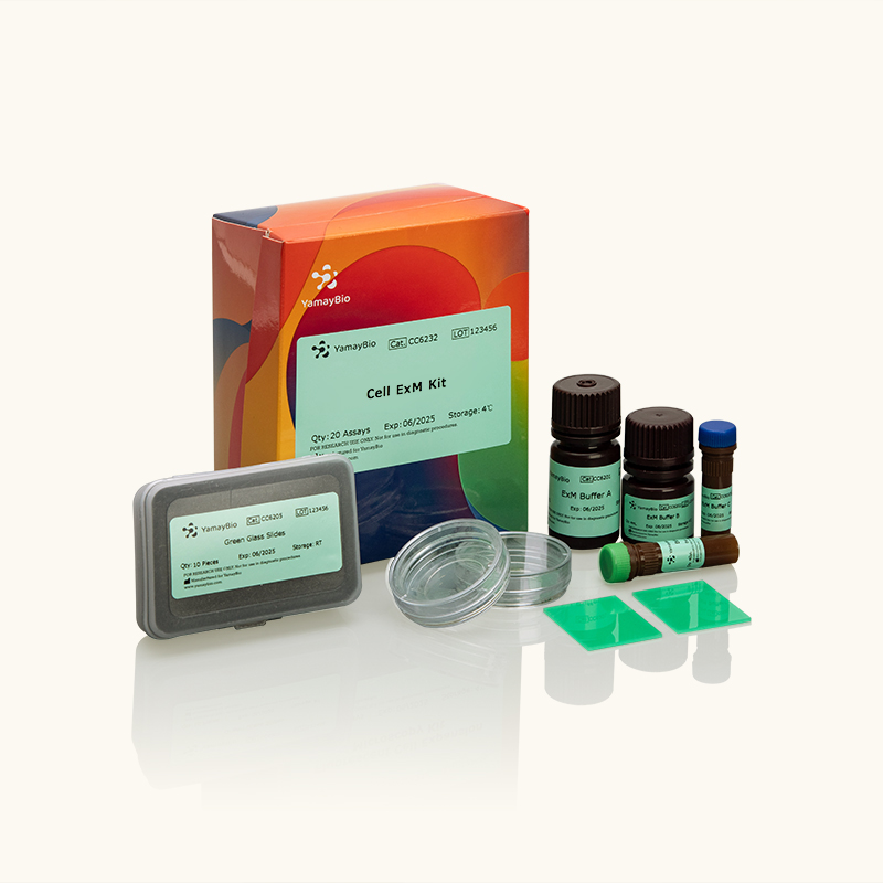


Cell ExM Kit
Expansion microscopy (ExM) is a novel super-resolution imaging technique. This technique is compatible with various biological molecules and can be implemented using wide-field and confocal microscopes. Read more
Transport at room temperature, store at 2~8℃, shelf life is 12 months; store at -20℃, shelf life is 36 months.
Expansion microscopy (ExM) is a novel super-resolution imaging technique. Biological samples can be isotropically expanded through expandable hydrogels, allowing for significantly improved resolution. This technique is compatible with various biological molecules and can be implemented using wide-field and confocal microscopes. For example, the resolution of wide-field microscopy can be improved from 250 nm to 60nm, and that of confocal microscopy from 120nm to 30nm. The uniform expansion preserves the spatial information of biomolecules.
KEY FEATURES

Significant Higher Imaging Resolution The image resolution was improved 4.5-fold in theory after expansion.

Preserves the Spatial Information of Biomolecules Uniform expansion preserves the spatial information of biomolecules.

Rapid and Efficient The cell sample experienced full volumetric expansion within 3 hours.
Hydrogel Expansion Fold in Three Dimension

Figure 1. Analysis of gel expansion fold .Gel was prepared by adding copper phthalocyanine at a concentration of 0.05%. Expanded gels demonstrated a 4.5-fold increase in linear dimensions (X,Y,Z). Dye intensity decreased following expansion due to dilution of dye molecules in the expanded hydrogel. Schematic representation of gel expansion in three dimension(a). Comparison of gel size before (left) and after (right) expansion in yz direction (b) and xy direction (c).
Significantly Increase Imaging Resolution
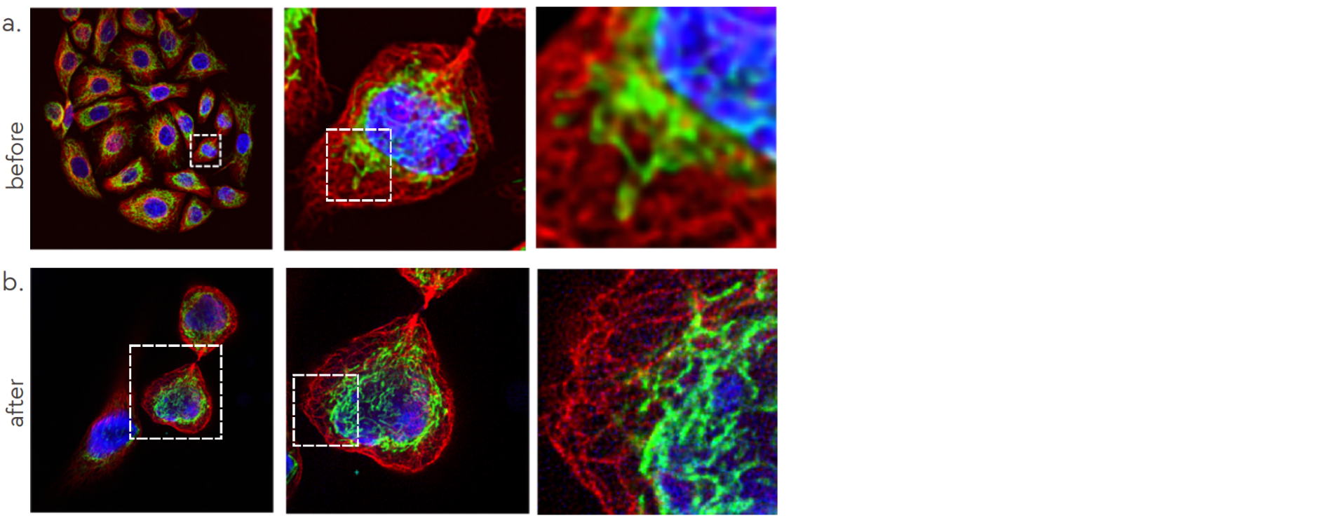
Figure 2. HeLa cells were imaged before (a) and after (b) expansion using a Nikon CSU-W1 confocal microscope followed by deconvolution with ImageJ. Hsp60 (green) labels mitochondria, tubulin (red) labels microtubules, and nuclei are stained with DAPI (blue).
The cell ExM kit would dramatically improve the imaging resolution of confocol microscopy.
Preserves the Spatial Relationship
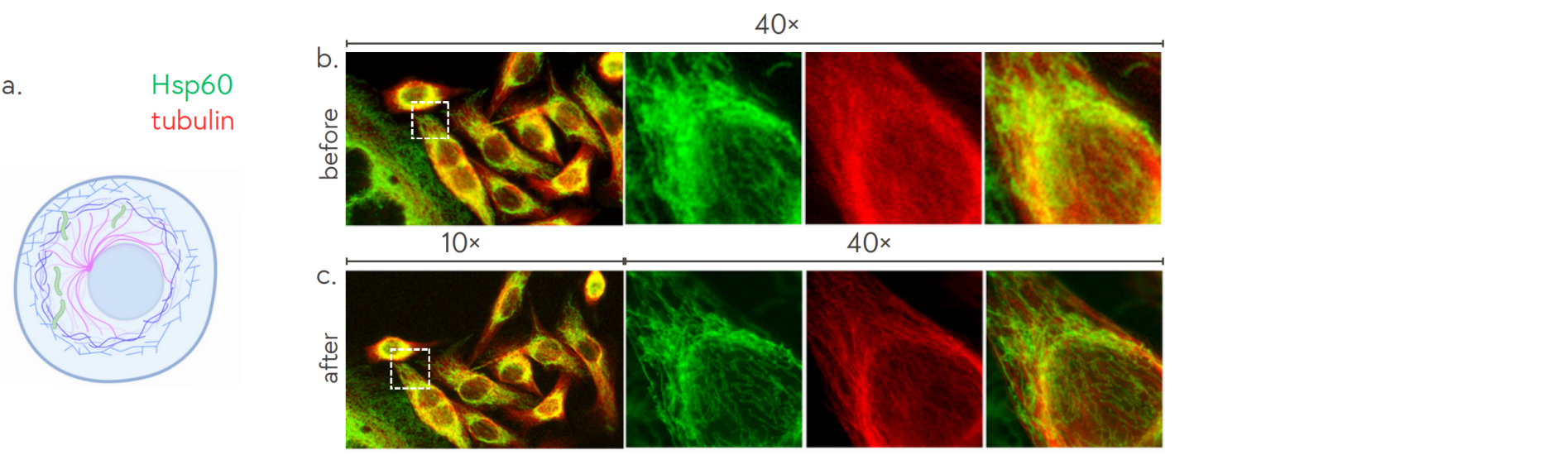
Figure 3. Schematic representation of subcellular localizaton of mitochondria and microtubles (a). HeLa cells were imaged before (b) and after (c) expansion using an OLYMPUS CKX53SF 40x microscope and subsequently deconvolved using ImageJ. Hsp60 (green) labels mitochondria, and tubulin (red) labels microtubules.
Enhanced resolution achieved through expansion microscopy revealed the original spatial relationships of proteins.
Traditionally, mitochondria and microtubules were not considered to exhibit significant colocalization.
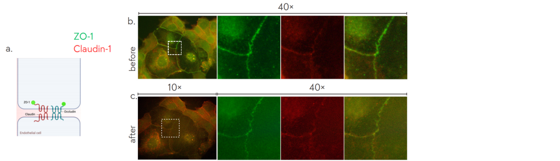
Figure 4. Schematic representation of cell-cell tight junction in Caco-2 cells (a). Caco-2 cells were imaged before (b) and after (c) expansion using an OLYMPUS CKX53SF 40x microscope and subsequently deconvolved using ImageJ. ZO-1 (green) and Claudin-1 (red) label cell-cell tight junctions.
ZO-1 and Claudin-1 are two well-established markers of cell-cell tight junctions, commonly used for analyzing protein-protein colocalization. Following expansion microscopy, improved resolution revealed more intricate details of the signal. Notably, signal intensity decreased due to the dilution of dye molecules within the expanded hydrogel.
Related Products


Certificate of Analysis


Safety Data Sheet
Product Manual
Specifications
| Application | Immunofluorescence (IF), FISH |
| Reagent Type | ExM Buffer A /ExM Buffer B/ExM Buffer C/ExM Buffer D/Green Glass Slides/Glass Bottom Dishes |
| Quantity | 1 Kit |
| Sufficient For | 20 Assays |